Digital fluoroscopy is a visualization method based on the use of X-rays. It allows for the real-time viewing of bone, soft tissue, and organ structures. In fluoroscopy, X-rays are captured by a special device that converts them, enhances their activity, and sends the resulting data to a computer. The computer processes the data and from multiple exposures of the object, a high-quality and dynamic real-time image is produced. This method enables the assessment of tissue and organ structures, their anatomical placement, as well as functionality. To conduct this diagnostics, patients are administered a high-density contrast agent to more precisely determine structural changes.
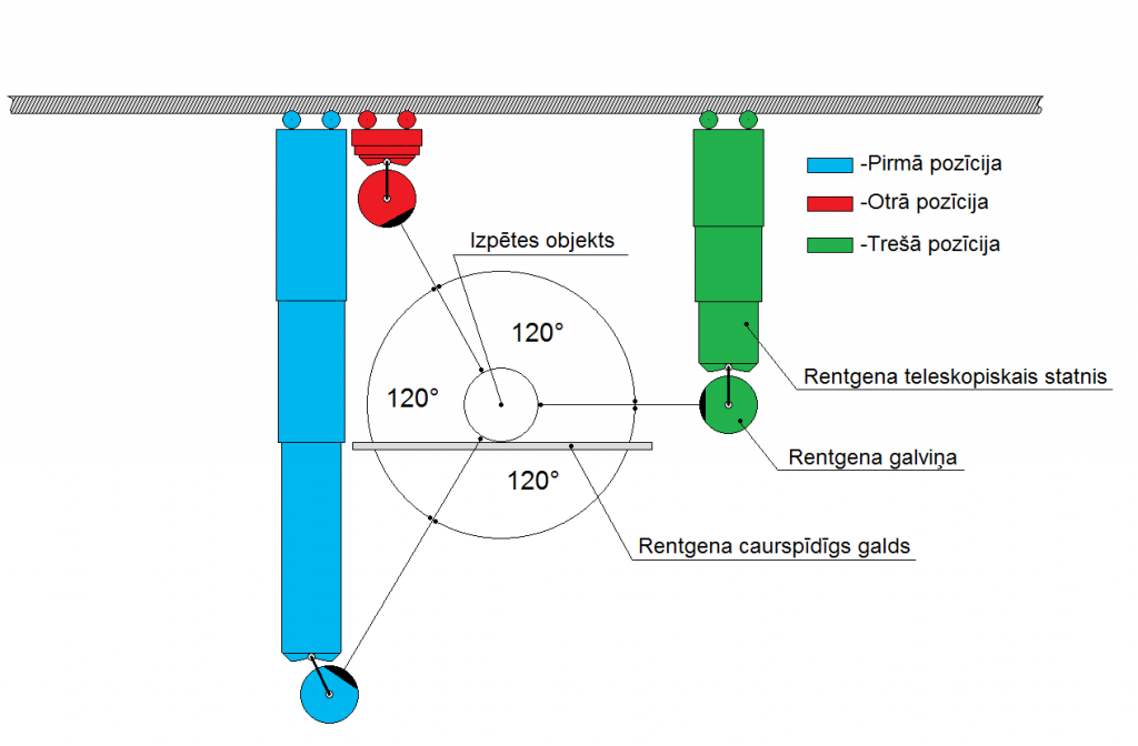
An X-ray tube with a collimator forms a conical beam of light that shines through the object. The X-rays, passing through the object, create an image that the computer captures. By rotating the object, many images are obtained which can be combined to create a 3D model of the object and its structure. Continuing with computer software, perspective distortions are removed. By scanning the illuminated object, many parallel images of the object are obtained. This is implemented using algorithms based on the inverse Radon transform. Using multiple X-ray integral absorption projections, these transforms allow for the reconstruction of a function that distributes density within the object. Finally, a set of specialized programs is applied, which allows viewing and converting many parallel images of the object and the 3D model.
This type of fluoroscopy is called digital fluoroscopy because the core data processing is performed with specialized software. Computed tomography also uses a special program, which in terms of cost is comparable to the entire X-ray apparatus.
Main clinical applications of the method:
– Barium sulfate examinations for digestive tract diagnostics: allows observation of how a patient swallows a barium sulfate solution and then tracks its movement through the digestive tract or performs an enema with barium.
– Hysterosalpingography: to assess the condition of the uterus and the patency of the fallopian tubes.
– Urethrogram and cystourethrogram during urination: to diagnose pathologies of the urinary tract and bladder.
– Fistulography: examination of fistulas.
– Repositioning of various bone fractures with visual control.
– Osteosynthesis and bone prosthetics for various pathologies.
– Detection of foreign bodies in the body, including those not visible in regular X-rays but can be visualized as a shadow.
– Determination of joint motion amplitude and mobility capabilities dynamically.
– Assessment of the extent of tumor infiltration at the site of localization.
Three-dimensional visualization of the tumor.

